Diseases/Disorders
Appendicitis
What is appendicitis?
Appendicitis is the inflammation of the appendix due to infection.
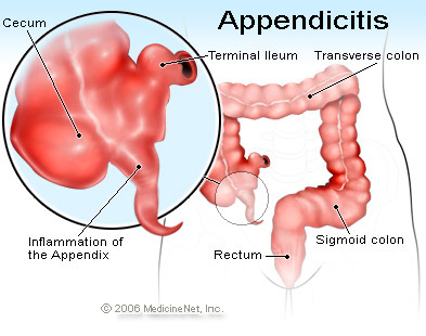
Where is the appendix located?
The appendix is a fingerlike pouch attached to the large intestine and found in the lower right of the abdomen.
Who is at risk for appendicitis?
Appendicitis occurs in both men and women usually between the ages of 15 and 30.
What are the causes of appendicitis?
The appendix is extremely small and is barely able to open. A foreign body or a mass of hardened feces could obstruct the entrance, so bacteria could multiply there causing inflammation.
What are the common symptoms of appendicitis?
Pain first occurs around the naval and the spreads towards the right side of the lower abdomen. The pain starts out centrally then pinpoints in one location. It is usually accompanied by nausea, vomiting, loss of appetite, constipation or diarrhea, inability to pass gas, a low-grade fever, and abdominal swelling. The abdominal pain occurs suddenly and increasingly grows worse. It is worse when moving around, coughing, sneezing, and taking deep breaths.
How is appendicitis diagnosed?
First the doctor will go over the patient's medical history in order to rule out other causes of the pain. Next they will do a physical examination where they will touch and apply pressure to the area where the patient is experiencing pain.
Reactions indicating appendicitis include:
Guarding- This is when a patient subconsciously tightens their abdominal muscles. This can occur voluntarily, which is when they tense their muscles right when the doctor's hand touches the abdomen. It could also be involuntary, which is when the patient's muscles tighten before the doctor's hand touches the abdomen.
Rebound Tenderness- This is when a doctor applies pressure to the abdomen and when the pressure is released pain occurs.
Rovsing's Sign- This is when pressure is applied to the lower left side of the abdomen and when the pressure is released pain occurs on the lower right side of the abdomen.
Psoa's Sign- The right psoa muscle over the pelvis is located near the appendix. Therefore when this muscle is used and the appendix is inflamed, pain will occur. A doctor tests for this by having the patient lie down and lift there right leg (with a bent knee) while the doctor adds resistance.
Obturator Sign- The obturator muscle is located close to the appendix, so when the appendix is inflamed and this muscle is used pain will occur. A doctor tests for this by having the patient lie down and with a bent knee move their right leg left and right.
Laboratory tests and imaging can also be done to diagnose an appendicitis. Blood tests can be used to see if there is infection. Computerized Tomography, x-rays, and ultrasounds can be used also. X-rays, however, are seldom helpful for diagnosing an appendicitis but can be used to find another explanation for the pain.
How is an appendicitis treated?
Usually, the appendix will be surgically removed in order to ensure that it won' burst. This is called an appendectomy. Nonsurgical treatment can be used, but research is still be conducted about this treatment, which would include antibiotics and a soft diet.
Why can the appendix be completely removed?
The appendix is a vestigial structure meaning that in an ancestral species the structure had a function but over time, but overtime the organism stopped using the structure. From scientist's understanding, the appendix does not have a function in the human body and therefore is not necessary for survival.
Appendicitis is the inflammation of the appendix due to infection.
Where is the appendix located?
The appendix is a fingerlike pouch attached to the large intestine and found in the lower right of the abdomen.
Who is at risk for appendicitis?
Appendicitis occurs in both men and women usually between the ages of 15 and 30.
What are the causes of appendicitis?
The appendix is extremely small and is barely able to open. A foreign body or a mass of hardened feces could obstruct the entrance, so bacteria could multiply there causing inflammation.
What are the common symptoms of appendicitis?
Pain first occurs around the naval and the spreads towards the right side of the lower abdomen. The pain starts out centrally then pinpoints in one location. It is usually accompanied by nausea, vomiting, loss of appetite, constipation or diarrhea, inability to pass gas, a low-grade fever, and abdominal swelling. The abdominal pain occurs suddenly and increasingly grows worse. It is worse when moving around, coughing, sneezing, and taking deep breaths.
How is appendicitis diagnosed?
First the doctor will go over the patient's medical history in order to rule out other causes of the pain. Next they will do a physical examination where they will touch and apply pressure to the area where the patient is experiencing pain.
Reactions indicating appendicitis include:
Guarding- This is when a patient subconsciously tightens their abdominal muscles. This can occur voluntarily, which is when they tense their muscles right when the doctor's hand touches the abdomen. It could also be involuntary, which is when the patient's muscles tighten before the doctor's hand touches the abdomen.
Rebound Tenderness- This is when a doctor applies pressure to the abdomen and when the pressure is released pain occurs.
Rovsing's Sign- This is when pressure is applied to the lower left side of the abdomen and when the pressure is released pain occurs on the lower right side of the abdomen.
Psoa's Sign- The right psoa muscle over the pelvis is located near the appendix. Therefore when this muscle is used and the appendix is inflamed, pain will occur. A doctor tests for this by having the patient lie down and lift there right leg (with a bent knee) while the doctor adds resistance.
Obturator Sign- The obturator muscle is located close to the appendix, so when the appendix is inflamed and this muscle is used pain will occur. A doctor tests for this by having the patient lie down and with a bent knee move their right leg left and right.
Laboratory tests and imaging can also be done to diagnose an appendicitis. Blood tests can be used to see if there is infection. Computerized Tomography, x-rays, and ultrasounds can be used also. X-rays, however, are seldom helpful for diagnosing an appendicitis but can be used to find another explanation for the pain.
How is an appendicitis treated?
Usually, the appendix will be surgically removed in order to ensure that it won' burst. This is called an appendectomy. Nonsurgical treatment can be used, but research is still be conducted about this treatment, which would include antibiotics and a soft diet.
Why can the appendix be completely removed?
The appendix is a vestigial structure meaning that in an ancestral species the structure had a function but over time, but overtime the organism stopped using the structure. From scientist's understanding, the appendix does not have a function in the human body and therefore is not necessary for survival.
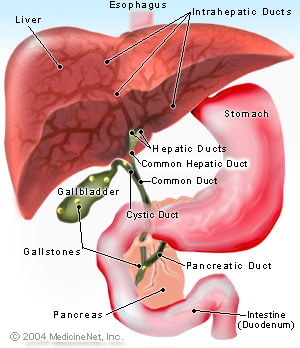
Gallstones
What are gallstones?
Gallstones are the accumulation of hardened cholesterol or calcium in the gall bladder.
Where is the gall bladder located?
The gall bladder is located just beneath the liver.
Who is at risk for gallstones?
Women are more at risk for gallstones than men especially if they are pregnant, use hormone replacements, or take birth control pills. People over age 60, American Indians, Mexican Americans, overweight or obese people, people who lose a lot of weight quickly or fast, people with a family history of gallstones, people with diabetes, and people who take cholesterol lowering drugs are at the most risk for gallstones also.
What are the causes of gallstones?
If the liver excretes more cholesterol than the bile can dissolve, the excess cholesterol could turn into stones. Another cause of gallstones is if the liver makes to much bilirubin, which is a factor in gallstone creation. Lastly, if the gallbladder doesn't empty properly or enough the build up of bile contributes to the development of gallstones.
What are the two types of gallstones that can be formed in the gall bladder?
The first type of gallstones are cholesterol gallstones, which are the most typical kind. They usually are yellow in color and are formed from undissolved cholesterol. The second type are pigment gallstones. These are black or brown in color and are formed when the bile has excess bilirubin.
What are the symptoms of gallstones?
A frequent symptom is pain in the upper stomach or in the upper right part underneath the ribs. The pain may start in the middle of the upper stomach, but then expand to the right upper back. The pain may last from 30 minutes to many hours and may occur after eating a meal. Vomiting could also occur as a result of the pain. Indications that the gallstone is obstructing the common bile duct include yellowing of the skin and whites of the eyes, dark urine, light colored stool, or/and a fever and chills.
How are gallstones diagnosed?
If gallstones are suspected, a doctor will most likely give the patient an ultrasound which would show if gallstones are present.
Other tests include:
Cholecystogram or Cholescintigraphy- A special iodine is injected into the patient and then after a certain amount of time an x-ray of the gall bladder is taken.
Endoscopic Retrograde Cholangiopancreatography- A doctor directs an endoscope through the stomach and into the small intestine of a patient. Then the doctor injects a dye that provisionally stains the biliary system's ducts. This allows the doctor to see where the gallstones are located.
Blood tests- Blood tests are used to test for infection, obstruction, or jaundice.
How are gallstones treated?
One solution to gallstones is to surgically remove the gall bladder. This is called a cholecystectomy. Nonsurgical treatment is used only when surgery is not an option for the patient. Oral dissolution therapy could be used. This is when drugs created from bile acid are used to dissolve gallstones. This option would take from months to years to dissolve all the gallstones. Contact dissolution is another type of therapy that is being experimented. This entails inserting a drug directly into the gallbladder to dissolve the gallstones. However, this has been found to have some complications.Why can a gall bladder be completely removed?Unlike the appendix, the gall bladder has a function, but people are able to live without it. This is because bile from the liver can move through the hepatic ducts into the common bile duct and right into the small intestine, instead of first storing the bile in the gallbladder.