Structure and Function of...
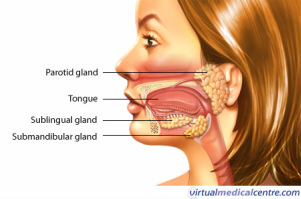
Oral Cavity (mouth)
The digestive tract starts with the oral cavity or in simpler terms the mouth. The oral cavity contains the accessory structures, the tongue, teeth, and salivary glands. There are three pairs of salivary glands, the parotid gland (on the masseter muscle below the ear), the submaxillary gland (a.k.a the submadibular gland, on the inside of the mandible), and the sublingual gland (in the floor of the mouth). As the teeth mechanically grind the food into small pieces the salivary glands produce saliva, which essentially activates the digestive process. The saliva lubricates the pieces of food, chemically breaks down carbohydrates with enzyme amerase, and also kills bacteria, so infections do not occur.
The digestive tract starts with the oral cavity or in simpler terms the mouth. The oral cavity contains the accessory structures, the tongue, teeth, and salivary glands. There are three pairs of salivary glands, the parotid gland (on the masseter muscle below the ear), the submaxillary gland (a.k.a the submadibular gland, on the inside of the mandible), and the sublingual gland (in the floor of the mouth). As the teeth mechanically grind the food into small pieces the salivary glands produce saliva, which essentially activates the digestive process. The saliva lubricates the pieces of food, chemically breaks down carbohydrates with enzyme amerase, and also kills bacteria, so infections do not occur.
Esophagus
The small food pieces travel from the oral cavity to the stomach through the esophagus. The esophagus is a 25 centimeter length tube of muscle, coated with a mucous membrane. The opening of this tube is able to expand, which it does when you swallow, but the rest of the length cannot so food must be small enough to pass through. The food mass which is now called a bolus moves down the esophagus by wave-like involuntary muscle contractions call peristalsis Digestive processes do not take place in the Esophagus, but it is necessary as a means of transport for the food.
The small food pieces travel from the oral cavity to the stomach through the esophagus. The esophagus is a 25 centimeter length tube of muscle, coated with a mucous membrane. The opening of this tube is able to expand, which it does when you swallow, but the rest of the length cannot so food must be small enough to pass through. The food mass which is now called a bolus moves down the esophagus by wave-like involuntary muscle contractions call peristalsis Digestive processes do not take place in the Esophagus, but it is necessary as a means of transport for the food.
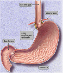
Stomach
The food is then stored in the stomach, combined with gastric juices, and broken down even more. The muscles on the walls of the stomach push the "food" together with wave-like movements (mechanical digestion) creating a thick semifluid mass called chyme. Gastric juice is produced from the glands in the gastric mucous membrane. This juice is made up of digestive juices, gastric acid, and gastric mucous. The digestive juices have digestive enzymes such as protease, which chemically digests proteins. The gastric mucous protects the membrane from the strong gastric acids. Gastric juices aid in the digestion of food, as well as eliminating pathogens that may have traveled to the stomach with the food. Stomach glands also secrete hydrochloric acid, which created the optimum PH for gastric protease and kills a lot of the bacteria remaining on food.
The food is then stored in the stomach, combined with gastric juices, and broken down even more. The muscles on the walls of the stomach push the "food" together with wave-like movements (mechanical digestion) creating a thick semifluid mass called chyme. Gastric juice is produced from the glands in the gastric mucous membrane. This juice is made up of digestive juices, gastric acid, and gastric mucous. The digestive juices have digestive enzymes such as protease, which chemically digests proteins. The gastric mucous protects the membrane from the strong gastric acids. Gastric juices aid in the digestion of food, as well as eliminating pathogens that may have traveled to the stomach with the food. Stomach glands also secrete hydrochloric acid, which created the optimum PH for gastric protease and kills a lot of the bacteria remaining on food.
Pancreas
The pancreas is an accessory organ located behind the stomach. It is made up of two different types of tissue. One type creates digestive juices, while the other makes the hormones insulin and glucagon (which regulate blood sugar levels). The digestive juices are produced in acini and enter the pancreatic duct. Then the juices leave through two openings of the duodenum (part of the small intestine). Proteins, carbohydrates, and fats in the duodenum are broken down by enzymes in the digestive juices.
The pancreas is an accessory organ located behind the stomach. It is made up of two different types of tissue. One type creates digestive juices, while the other makes the hormones insulin and glucagon (which regulate blood sugar levels). The digestive juices are produced in acini and enter the pancreatic duct. Then the juices leave through two openings of the duodenum (part of the small intestine). Proteins, carbohydrates, and fats in the duodenum are broken down by enzymes in the digestive juices.
Liver and Gall Bladder
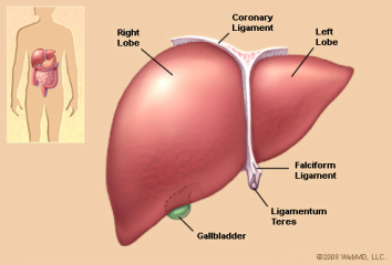
The liver and gall bladder are both accessory organs. The liver is located beneath the diaphragm and the gall bladder (small pear shaped sack) is hidden below the liver. The liver takes toxins out of the blood and makes bile that aids the body in absorbing fat. The gall bladder is used to keep the bile until it is needed. When it is needed bile moves through ducts to enter the small intestine, where it will be used to help break down food.
The liver and gall bladder are both accessory organs. The liver is located beneath the diaphragm and the gall bladder (small pear shaped sack) is hidden below the liver. The liver takes toxins out of the blood and makes bile that aids the body in absorbing fat. The gall bladder is used to keep the bile until it is needed. When it is needed bile moves through ducts to enter the small intestine, where it will be used to help break down food.
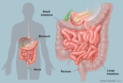
Small Intestine
From the stomach, the Chyme travels to the small intestine. The interior of the small intestine is coated with a mucous membrane, which has an increased surface area from the many folds and villi. The mucous membrane produces digestive juices that finish the digestive process with the help of the digestive juices from the pancreas and the bile. This part of the process mostly occurs in the duodenum, a portion of the small intestine. The key job of the small intestine is to chemically break down the remaining undigested parts of the chyme and absorb the nutrients from it into the blood. The villi absorb the nutrients in the jejunum and ileum, other sections of the small intestine. The nutrients then enter the bloodstream.
From the stomach, the Chyme travels to the small intestine. The interior of the small intestine is coated with a mucous membrane, which has an increased surface area from the many folds and villi. The mucous membrane produces digestive juices that finish the digestive process with the help of the digestive juices from the pancreas and the bile. This part of the process mostly occurs in the duodenum, a portion of the small intestine. The key job of the small intestine is to chemically break down the remaining undigested parts of the chyme and absorb the nutrients from it into the blood. The villi absorb the nutrients in the jejunum and ileum, other sections of the small intestine. The nutrients then enter the bloodstream.
Large intestine
The ileocecal valves of the small intestine passes undigestible wastes to the large intestine. The large intestine reabsorbs water and is responsible for egestion, which is the elimination of solid wastes. Wastes are moved through the ascending, transverse, descending, and sigmoid portions of the colon by peristalsis. As they move along water from them is absorbed. Since water is removed from the wastes they become more solid as they near the rectum (lower end of large intestine). The wastes, which are compressed into feces, are stored in the rectum until elimination, which occurs through the anus.
The ileocecal valves of the small intestine passes undigestible wastes to the large intestine. The large intestine reabsorbs water and is responsible for egestion, which is the elimination of solid wastes. Wastes are moved through the ascending, transverse, descending, and sigmoid portions of the colon by peristalsis. As they move along water from them is absorbed. Since water is removed from the wastes they become more solid as they near the rectum (lower end of large intestine). The wastes, which are compressed into feces, are stored in the rectum until elimination, which occurs through the anus.